
- Asthma is a chronic respiratory disease characterized by airway obstruction (narrowing of airways), difficulty in breathing, wheezing and shortness of breath which may be triggered by exposure to certain allergens, stress, exercise etc.
- Different anti-asthmatic drugs are available in market. Some of them are:
- Beta adrenergic agonists (e.g., Salbutamol, salmeterol)
- Corticosteroids (e.g., Hydrocortisone, beclomethasone)
- Leukotriene Modifiers (Montelukast, Zafirlukast).
- There are different methods of screening of anti-asthmatic agents which are mentioned and explained below.
Screening of Anti-asthmatic agents
1) In-vitro methods for screening of anti-asthmatic agents
Histamine (H1) receptor binding
2) Effect on airways
We can use in-vivo methods or perform test in isolated organs to find effect on airways.
Tests in isolated organs
- Spasmolytic activity in isolated trachea
- Spasmolytic activity in isolated guinea pig lung strips
- Bronchial perfusion of isolated lung
- Vascular and airway response in isolated lung
- Reactivity of isolated perfused trachea
In-vivo methods for screening of anti-asthmatic agents
- Bronchial hyperreactivity
- Broncho spasmolytic activity in anesthetized guinea pig (Konzett-Rossler method)
- Effect of arachidonic acid or PAF in respiratory function
- Airway microvascular leakage
- Pneumotachography in anesthetized guinea pigs
3) Anti-tussive Activity
4) Effect on tracheal cells, bronchial mucous secretion, and transport
Broncho spasmolytic activity in anesthetized guinea pig (Konzett- Rossler method)
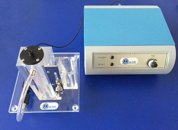
Figure – Bronchospasm transducer which can be used to access airflow resistance while screening of anti-asthmatic agents (source- animalab.eu)
Purpose and rationale
- Konzett and Rossler described this method which is based on measurement of air volume changes in living animal. Spasmogens like histamine, PAV, bradykinin etc. cause bronchospasm during which volume of inspired air decreases and of excess air increases.
- In this method, drug’s broncho spasmolytic activity is measured by measuring volume of air which is not taken by lungs after bronchospasm. The inhibition of bronchospasm produced by test substance and standard is measured and compared.
- It is one of the widely accepted screening model of anti-asthmatic.
Animals used
- Guinea pig of either sex weighing around 250-500 gm.
Procedure
- Suitable anesthetic agent is used to anesthetize guinea pig. After this trachea is cannulated by two-way cannula. One arm of cannula is connected to respiratory pump via which guinea pig is artificially respired and another arm is connected to Statham P23 Db transducer.
- Spasmogens and test substance or standard is administered via cannula in interna jugular vein. Excess air which is not taken by lungs is measured by polygraph.
- Suitable spasmogen is administered via IV route and look for induction of bronchospasm. After two bronchospasm of equal intensity, test compound is administered by suitable route (i.v., p.o., subcutaneous or intraduodenal route).
- Spasmogen is administered again, and the interval of administration depend on route of test drug.
- 5,15 and 30 minutes after i.v. administration of drug.
- 15, 30 and 60 minutes after intraduodenal administration of drug and.
- 30 and 60 minutes after oral administration of drug.
- Standard drug used depend on spasmogenic agent used. For e.g.:
- Aminophylline for bradykinin induced spasm.
- Atropine sulphate for methacholine or acetylcholine induced spasm.
Evaluation
- Percent inhibition of induced bronchospasm is measured for test substance and compared with control. ED50 value is calculated.
Bronchial hyperreactivity
Purpose and rationale
- Inhalation of spasmogens like histamine produce symptoms like forced inspiration, increased breathing frequency and asphytic convulsions which resemble asthma. Use of drugs delay occurrence of these symptoms.
- In this method, pre-convulsion time (time until asphytic convulsion) is measured.
Animals used
- Male guinea pig weighing around 300-400 gm. Each group consist of 10 guinea pigs.
Device used
- Devised used is inhalation cage which consist of 3 boxes- A, B and C and all of them are well- ventilated.
Procedure
- Animals are first kept in box A where test drug or standard drug is applied by ultra-sound nebulizer or by oral or sc route.
- After administering the drug, they are passed to box C through box B. In box C, aerosol of 0.1% histamine HCl is applied by ultra-sound nebulizer. Animals are observed and time until first asphytic convulsion is noted.
- After the guinea pig experience first asphytic convulsion, they need to be immediately removed from box C.
Evaluation
- Pre-convulsion time is measured for both test substance and standard and compared. ED50 value is calculated.
References
- Patil SD et al. In-vivo and in-vitro screening models of asthma: an overview. IJRDPL. June – July, 2016: 5(4): 2209-2218.
- MS Shreedevi etal. Various Screening Methods for Anti- Asthmatic Activity. Current Traditional Medicine. 2015: 1(2); 108-15.
- Drug Discovery and Evaluation: Pharmacological Assay. 2nd edition.
- Lippincott’s Illustrated Reviews Pharmacology. 6th edition.
- https://pubmed.ncbi.nlm.nih.gov/10893704/