Anatomy of Skin
• Skin is the largest organ of body, comprising about 15 % of total body weight of an adult. It is one of the most complicated, ever-changing organs and contains many specialized cells and structures.
• The skin and its accessory structures make up the integumentary system which provides the body with overall protection.
• The three major layers of skin are epidermis, dermis and subcutaneous tissue or hypodermis.
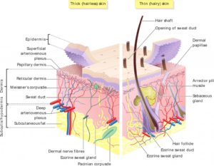
Figure 1- A section of skin (Source- Madhero88, via Wikimedia Commons, CC BY-SA 3.0 License)
Epidermis
• The epidermis is outermost among three layers of skin and is composed of keratinized, stratified squamous epithelium. It is made of four or five layers of epithelial cell, depending on its location of the body.
• Most abundant two types of cells present are keratinocytes and dendritic cells. There are no capillaries present between them (it is avascular).
• Skin that has four layers of cell is known as ‘thin skin’. These four layers are stratum basale, stratum spinosum, stratum granulosum, and stratum corneum. Most of the skin can be classified as thin skin. E.g. it is thinnest on the eyelids (half a millimeter). “Thick skin” has a fifth layer, called the stratum lucidum, located between the stratum corneum and the stratum granulosum. It is found only on the palms and soles (1.5 millimeter).
• It regenerates continuously throughout the life. It is estimated that epidermis turns over every 50-56 days in human.

Figure 2- Different layers of epidermis and dermis
Stratum basale
• This layer is also known as stratum germinativum. The name germinativum is due to its capacity to divide or germinate and the name basale is due to its lowest position or base.
• It is the deepest epidermal layer and has column shaped basal cells that divide and push older cells toward the surface of the skin. As the cells move up, they flatten up, die and shed. These cells produce the protein ‘keratin’.
• The cells in stratum basale are connected to dermis though intertwining collagen fibers known as basement membrane. This layer also contains melanin transferred from adjoining melanocytes.
Stratum spinosum
• This is the thickest layer of the epidermis. It is composed of 8-10 layers of keratinocytes, which are strengthening proteins, formed as a result of cell division in stratum basale. It also contains Langerhans cells (a type of dendritic cells), that help to prevent infection by acting as macrophage.
• It is spiny in appearance due to protruding cell processes that joins the cells via a structure called desmosomes.
Stratum granulosum
• It is also known as granular layer and consist of stratified squamous cells arranged in 1-3 layers. It consists of lamellar granules which is made up of fibrous keratohyalin.
• The two proteins keratin and keratohyalin produced by keratinocytes give grainy appearance. As the cells die, the nuclei and other cell organelles disintegrate and leave behind keratin, keratohyalin and cell membrane which form stratum corneum, stratum lucidum and accessory structures of hair and nails.
Stratum lucidum
• This layer is found only in thick skin of palm, soles and digits between the stratum corneum and stratum granulosum.
• It is a smooth, translucent layer of epidermis which consist of dead and flattened keratinocytes. These cells are densely packed with eleidin (a clear protein rich in lipids) which gives lucid appearance to this layer. Hence the name lucidum is given.
• It functions as a barrier and has waterproof properties.
Stratum corneum
• It is the outermost epidermal layer and is the layer exposed to outer environment. It is also known as horny cell layer. It consists mainly of keratinocytes containing a protein known as keratin.
• The name ‘corneum’ is due to excessive keratinization or cornification of the cells in this layer.
• There are about 15-30 layers of cell. Cells in this layer are shed periodically and is replaced by new cells from stratum basale. The entire layer is replaced during a period of about 4 weeks.
• This layer acts as barrier to various pathogens and chemicals. It provides mechanical protection against abrasion and prevent dehydration of underlying tissues. Its importance become apparent when it is lost (like during burns).
Dermis
• It is an integrated system of fibrous, filamentous and amorphous connective tissue. There are two layers of connective tissue that compose interconnected mesh of elastin and collagenous fibers produced by fibroblast.
• The two main characteristics of this layer are strength and elasticity. As the age increases, the skin loses its elasticity due to deterioration of elastin fibers. Hence, wrinkling occurs.
• It can be divided into two layers; the papillary dermis which is thin, upper layer and the reticular dermis, which is thick, lower layer.
• It contains specialized cells and structures including;
– Hair follicles.
– Sebaceous glands (produces an oily substance called sebum).
– Blood vessels and nerve endings.
– Apocrine and endocrine glands.
– Meissner corpuscles and laminar corpuscles that transmit sensation of touch and pain.
• Its main role is to make sweat and oil, provide sensations and blood to the skin and grow hair.
Subcutaneous tissue
• It is also known as superficial fascia, hypodermis or subcutis. It helps in connecting skin with underlying fascia (fibrous tissue) of bones and muscles. Technically, it may not be considered as a part of skin.
• It mainly consists of well vascularized, loose, areolar connective tissue and adipose tissue which help in storage of fat. This fat layer helps to avoid excess heat loss and protect bones and muscles.
• It helps in providing main structural support to skin, insulate body from cold and aid in shock absorption.
Functions of the skin
Protection and repair
• Its primary function is to serve as protective barrier from mechanical, thermal and other physical injury, harmful agents and excessive loss of moisture and protein. Melanocytes provide protection from harmful UV rays.
Thermoregulation
• It helps to maintain constant core temperature and protect the body from cold or heat. This is achieved by alteration of blood flow through the cutaneous vascular bed.
• The secretion and evaporation of sweet from the body also helps to cool the body.
Sensation
• It is sense-of-touch organ which triggers a response when we touch or feel something, including things that may cause pain. The nerve endings present help in detecting temperature, vibration, pressure, touch and injury.
Immunity
• Langerhans cells in skin are part of immune system. Langerhans cells and dendritic cells takes up microbial present in skin to transform into antigen presenting cells and provide immunity by interacting with T cells.
Storage
• It stores fat to provide insulation.
Biochemical functions
• It is involved in various biochemical processes like synthesis of vitamin D in the presence of sunlight. Vitamin D is essential for normal calcium and phosphorus absorption required for healthy bones.
References
- Paul AJ, Ann M, Carolyn G. Anatomy and Physiology of the Skin. Journal of the Dermatology Nurses’ Association. 2011; 3(4): 203-213.
- Koster MI. Making an epidermis. Ann N Y Acad Sci. 2009; 1170: 7–10.
- Byrd AL, Belkaid Y, Segre JA. The human skin microbiome. Nature Reviews Microbiology. 2018; 16: 143–155.
- Essentials of Anatomy and Physiology. 5th edition.