• G-protein coupled receptors or GPCR are large group of transmembrane receptors that act as molecular switches by converting between on and off state and convert extracellular signal to intracellular signal. The name G-protein coupled receptor (GPCR) is given as they are linked to G proteins (guanine nucleotide binding proteins). GPCR are also known as seven transmembrane receptor or heptahelical receptor. It is because its structure consists of a single polypeptide chain that threads back and forth 7 times through lipid bilayer of plasma membrane. The existence of GPCR was demonstrated by Robert J. Lefkowitz- an American physician and molecular biologist. He shared the 2012 Nobel prize for Chemistry with his colleague Brian Kobilka for elucidation in GPCR structure and function.
• Some examples of G-protein coupled receptor are dopamine receptors, adrenergic β1 and α2 receptors.
Location of G-protein Coupled Receptor
• They are found in cell membrane of wide range of organisms including mammals, microorganisms and plants. In human body, they are found on all cell types and are abundant in brain and gut.
Structure of G-protein coupled receptor
• GPCR is made up of long protein that consist of 3 basic regions; an extracellular portion (the N terminus), an intracellular portion (the C terminus) and a middle segment which consist of 7 membrane-spanning domains or transmembrane helices.
• The long middle segment transverse the membrane seven times in a serpentine pattern.
G-protein
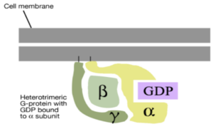
Figure 1- Inactive G-proteins with bound GDP
• G-proteins also known as guanine nucleotide binding protein are linked to GPCR. They are associated with cytosolic side of plasma membrane.
• G-proteins can be classified into two types; monomeric small GTPases (small G proteins) and heterotrimeric G protein complexes. Heterotrimeric G protein are composed of 3 subunits, alpha (Gsα), beta (Gβ) and gamma (Gϒ). So, the name heterotrimeric (hetero- different, trimeric- having 3 parts).
• G-proteins can bind to and hydrolyze GTP (Guanosine Triphosphate) to GDP (guanosine di-phosphate). They are ‘on’ when bound to GTP and ‘off’ when bound to GDP.
How G-protein coupled receptor (GPCR) work?

Figure 2– Mechanism of action of of G-protein coupled receptor (source- G protein” By Tpirojsi – Own work (Public Domain) via Commons Wikimedia)
• The activation of GPCR requires binding of agonist. This agonist may be a hormone, neurotransmitter or an external stimulus such as pheromone or odor. The binding of agonist to GPCR initiate a series of reactions which trigger a cellular response.
1. When GPCR is not bound to an agonist, it remains inactive and G-protein also remain inactive on the cell membrane. The GDP is bound to Gsα domain of G-protein.
2. When agonist binds to GPCR, it undergoes a conformational change and activate its GEF domain. This change in confirmation allows binding of G-proteins to GEF domain and GDP of G-protein is replaced by GTP leading to activation of G-proteins.
3. After activation Gsα domain gets dissociated from GPCR- G protein complex and binds to effector enzyme (adenylyl cyclase, phospholipase C) present on cell membrane to activate it. This generates second messenger such as cAMP, inositol 1,4,5- triphosphate, 1,2-diacylglycerol etc. which activate various proteins present in cytosol and generate particular cell response.
4. The hydrolysis of GTP into GDP in Gsα domain dissociate it from effector enzyme resulting in inactivation of enzyme.
Classification of G-protein coupled receptor
GPCR can be classified into 6 types (A-F) based on sequence homology and functional similarity. The 6 types are; Class A or 1- rhodopsin like, Class B or 2- secretin like, Class C or 3- metabotropic glutamate, Class D or 4- pheromone which are found on fungal species, Class E or 5- Cyclic AMP receptors expressed exclusively on unicellular amoeba and Class F or 6- frizzled or smoothened receptors.
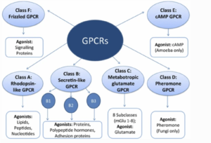
Figure 3- GPCRDB database classification of G-protein coupled Receptor (GPCR) based on agonist affinity and functions (Source- Moran et al, 2016)
Physiological Role of GPCR
There are more than 1000 known GPCR in human body each having different function. Various physiological roles of GPCR include:
• Regulation of immune system activity and inflammation.
• Regulation of sympathetic and parasympathetic nervous system and helps in controlling many autonomic functions like digestive process, heart rate and blood pressure.
• Homeostasis modulation.
• Behavioral and mood regulation.
• For visual sense, sense of smell and gustatory sense.
Pharmacological Role of GPCR
• Disruption of GPCR signaling is associated with many diseases including diabetes, allergies, some cancer and some cardiovascular disease. So, it is target of many modern drugs.
• Some example of drugs which target GPCR are;
– Platelet directed therapeutics like clopidogrel and ticlopidine which target P2Y12 receptor for adenosine diphosphate (ADP).
– Many drugs used in asthma like terbutaline, salmeterol and formoterol bind to GPCR at β2 receptor and act as bronchodilator.
– Cholinergic agonist like carbachol and pilocarpine which are used to treat glaucoma, bind to GPCR and produce their pharmacological actions.
– Antipsychotics (old generation like chlorpromazine, haloperidol as well as new generation like risperidone, quetiapine) bind to GPCR at dopaminergic pathways to decrease their hyper functionality.
– Opioids bind to GPCR of opioid receptor.
Reference
- https://en.wikipedia.org/wiki/G_protein
- https://egpat.com/blog/15-important-gpcr-binding-drugs-in-pharmacology
- Moran BM, Flatt PR, McKillop AM. G protein-coupled receptors: signalling and regulation by lipid agonists for improved glucose homoeostasis. Acta Diabetologica. 2016; 53: 177-188.
- Woulfe DS. REVIEW ARTICLES: Platelet G protein‐coupled receptors in hemostasis and thrombosis. jth. 2005; 3(10): 2193-2200.
- Wendell SG, Fan H, Zhang C. G Protein–Coupled Receptors in Asthma Therapy: Pharmacology and Drug Action. Pharmacological Reviews. 2020; 72 (1): 1-49.
Excellent effort
Thank you. Please, keep visiting our website.